TL;DR: Mount Sinai researchers showed APOE4 actively converts brain pericytes into scar-forming cells and disrupts astrocyte waste clearance, and blocking TGF-β signaling reversed vascular damage in lab models.
On September 24, 2026, researchers at the Icahn School of Medicine at Mount Sinai reported that APOE4, the strongest genetic risk factor for Alzheimer's disease, damages brain blood vessels through an active and potentially reversible biological process, according to the Mount Sinai news release titled Mount Sinai Researchers Identify How APOE4 Gene Damages Brain Blood Vessels in Alzheimer's Disease.
The work was published as two companion studies in the journals Cell and Cell Stem Cell, according to that Mount Sinai announcement. Both studies used human brain tissue and miBrains, a stem cell-derived human brain tissue platform, to trace how APOE4 promotes vascular scarring and abnormal protein buildup.
Alzheimer's disease is a progressive brain disorder that gradually impairs memory, thinking, and behavior, according to the same announcement. It affects more than 7 million older adults in the United States. Scientists have long known that the brain's blood vessels deteriorate during the disease, particularly in people carrying APOE4, but the underlying mechanisms had remained unclear.
That gap matters because cerebrovascular damage has often been viewed as a downstream consequence of Alzheimer's disease rather than a process that actively contributes to disease progression, according to the Mount Sinai announcement. The new studies reframe vessel injury as a biologically active process caused by APOE4 that may be reversible.
Two studies traced APOE4 damage in vessels and brain tissue
The Mount Sinai team examined both blood-vessel degeneration and toxic protein accumulation, according to the Mount Sinai announcement. One study published in Cell focused on the brain vasculature. A second study published in Cell Stem Cell focused on buildup of abnormal proteins associated with neurodegenerative disease.
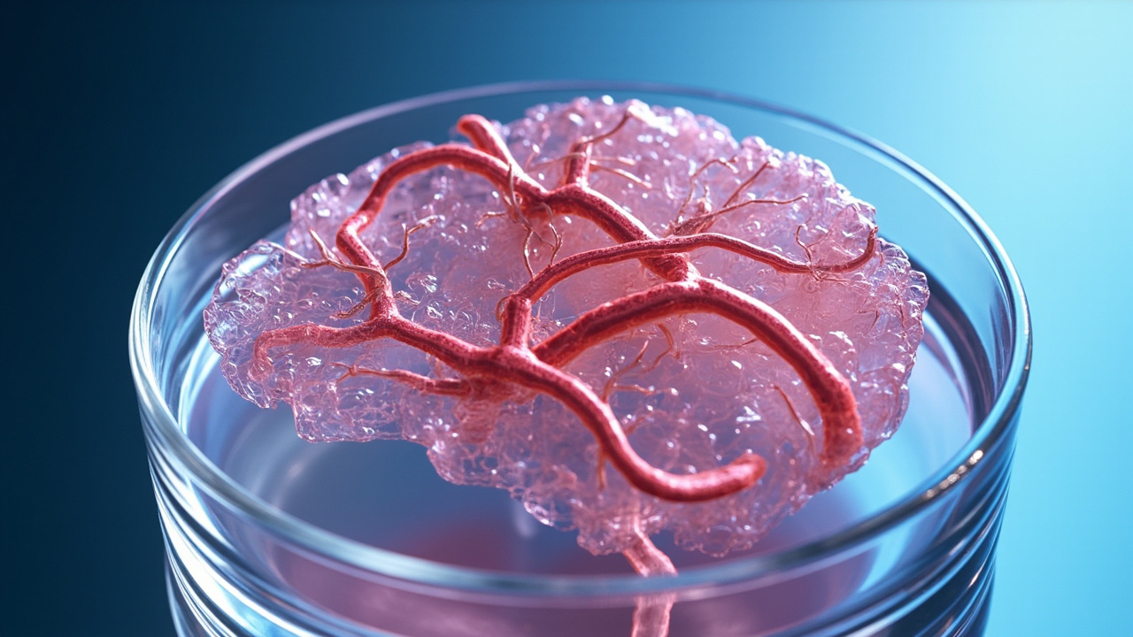
Both studies used miBrains, three-dimensional human brain tissue developed by the Mount Sinai team and derived from induced pluripotent stem cells, according to the same announcement. The platform was designed to model key features of human brain tissue and its vascular network.
The Blanchard laboratory integrated preclinical models, postmortem human brain tissue, transcriptomics data, and miBrains, according to the Mount Sinai announcement. Each system validated and extended findings from the others. The researchers said this approach made it possible to reconstruct events leading to end-stage vascular pathologies seen in postmortem human brain tissue, define mechanisms, and test candidate interventions.
A vascular atlas mapped gene activity cell by cell
In the Cell study published on September 24, Mount Sinai researchers combined existing datasets to create a single-cell transcriptomic atlas of the human brain's vasculature, according to the Mount Sinai announcement. The atlas provided a detailed map of gene activity across cells that form and support blood vessels.
The paper, titled A pericyte-to-myofibroblast transition links APOE4 to cerebrovascular degeneration, identifies an APOE4-enriched myofibroblast population and reports that TGF-β inhibition reverses the transition, according to the Cell article summary. The study was received September 3, 2025, revised March 20, 2026, accepted August 28, 2026, and published online September 24, 2026.
According to the Cell summary, the authors assembled a single-cell transcriptomic atlas of human brain vasculature to uncover how APOE4 promotes cerebrovascular pathology. In APOE4 carriers, pericyte abundance was significantly reduced and accompanied by emergence of a myofibroblast-like cell population co-expressing contraction and extracellular matrix genes.
Immunostaining confirmed non-vascular myofibroblasts in APOE4 human and mouse brains, according to the same Cell summary. The authors reported that APOE4 pericytes transition into myofibroblasts that secrete fibronectin, which promotes vascular amyloid accumulation.
Pericytes converted into scar-producing cells
Pericytes are cells that normally stabilize small blood vessels and help maintain the blood-brain barrier, according to the Mount Sinai announcement. The Cell study found that APOE4 caused those cells to transform into scar-forming myofibroblast-like cells.
This transition promoted vascular fibrosis and increased amyloid accumulation around the vessels, according to the same announcement. Those changes create conditions that may compromise blood flow and promote neurodegeneration.
The Cell article summary describes the same sequence as a pericyte-to-myofibroblast transition. It states that elevated transforming growth factor β signaling was identified as the driver of the transition through computational and experimental analyses. Excessive fibronectin deposition occurred along the vasculature, and fibronectin secretion from APOE4 myofibroblasts directly promoted perivascular amyloid accumulation.
As summarized by Neuroscience News in APOE4 Damages Brain Vessels and Halts Waste Disposal, the analysis showed APOE4 prompts pericytes to undergo pathological transdifferentiation into scar-producing cells. That report states the transformation drives vascular fibrosis and accelerates deposition of amyloid-beta around cerebral vessels.
Blocking TGF-β restored vessel coverage in models
Importantly, blocking TGF-β signaling, a pathway involved in cellular communication and tissue remodeling, restored pericyte coverage and reduced both fibrosis and vascular amyloid, according to the Mount Sinai announcement. The team reproduced the finding in aged APOE4 mice, showing that APOE4-associated cerebrovascular degeneration can be therapeutically reversed in that model.
According to the Cell summary, inhibition of TGF-β restored pericyte coverage and reduced vascular fibrosis and amyloid to APOE3 levels. The authors described the result as revealing a targetable mechanism linking APOE4 to cerebrovascular pathology in Alzheimer's disease.
Neuroscience News, summarizing the same studies, reported that inhibiting the TGF-β signaling pathway reversed the transition. In aged APOE4 animal models, blocking TGF-β restored pericyte coverage, suppressed fibrotic scarring, and significantly diminished amyloid accumulation around blood vessels.
Corresponding author Joel W. Blanchard, PhD, Associate Professor of Neuroscience, and Stem Cell Biology and Regenerative Medicine at the Icahn School of Medicine at Mount Sinai, said vessel damage is not simply a late consequence of disease, according to the Mount Sinai announcement.
"Damage to the brain's blood vessels is not simply a late consequence of Alzheimer's disease; it is a biologically active process caused by APOE4 that may be reversible," Blanchard said.
He added that the findings reveal new therapeutic targets for preserving vascular function and limiting amyloid accumulation. The statement was reported in the Mount Sinai announcement and repeated in coverage by Neuroscience News.
First author Braxton R. Schuldt, MD/PhD candidate in Neuroscience and researcher in the Blanchard Laboratory, said the experiments showed conversion of vessel-support cells into scar-producing cells and accumulation of amyloid around vessels, according to the same announcement.
"We show that APOE4 converts blood-vessel support cells into scar-producing cells, causing amyloid or abnormal protein buildup to accumulate around the brain's vessels," Schuldt said. "Through our experiments, we were able to block this protein buildup process, revealing possible new therapeutic treatment options and strategies for protecting the brain's circulation in people at high genetic risk for Alzheimer's disease."
Second study examined toxic protein buildup
In the Cell Stem Cell study, the Blanchard laboratory used miBrains to investigate how APOE4 promotes buildup of abnormal proteins in neurodegenerative disease, according to the Mount Sinai announcement. Such protein accumulation is a hallmark of disorders including Alzheimer's and Parkinson's disease.
Studying how protein accumulation develops in the living human brain is exceptionally difficult, according to the same announcement. The miBrain system provided a window into otherwise inaccessible processes in complex, human brain-like tissue.
MiBrains contain all the major cell types found in the human brain, including neurons, supporting glial cells, myelin-producing cells, and cells that form blood vessels, according to the Mount Sinai announcement. Like the human brain, miBrains carrying APOE4 accumulate higher levels of abnormal alpha-synuclein, a protein most commonly associated with Lewy body dementia and Parkinson's disease.
Despite its broad clinical significance, the cellular mechanisms that cause alpha-synuclein to accumulate have been poorly understood, according to the same announcement. The Cell Stem Cell experiments addressed that question by tracking cholesterol, lysosomal function, and protein clearance in human-like tissue.
APOE4 disrupted astrocytes and waste disposal
Experiments showed that APOE4 causes cholesterol to accumulate in astrocytes, support cells with essential roles in maintaining brain health, according to the Mount Sinai announcement. This excess cholesterol impairs the astrocytes' lysosomal waste-disposal system, reducing their ability to break down alpha-synuclein.
The protein instead aggregates and spreads to neurons, leading to harmful protein deposits, according to the same announcement. The findings identify cholesterol metabolism in astrocytes and lysosomal function as promising therapeutic targets for Alzheimer's and Parkinson's disease.
Neuroscience News described the same cascade as cholesterol congestion followed by lysosomal breakdown and pathological spread. In that summary, APOE4 triggers abnormal cholesterol storage inside astrocytes, disrupts lysosomal machinery, and allows toxic alpha-synuclein to build up, aggregate, and spread into neighboring neurons.
The Mount Sinai announcement states that researchers identified a chain of events linking APOE4 to lipid accumulation in astrocytes, impaired clearance of alpha-synuclein, and toxic protein deposits. That chain points to lipid metabolism and cellular waste-removal pathways as potential therapeutic targets.
miBrains supplied neurons, glia and vessels in one model
The studies relied on miBrains to bring multiple brain cell types together in three-dimensional tissue, according to the Mount Sinai announcement. The model included neurons, supporting glial cells, myelin-producing cells, and cells that form blood vessels.
Because the tissue is derived from induced pluripotent stem cells, researchers can introduce defined genetic factors such as APOE4 and observe effects in human-like tissue, according to the same announcement. Neuroscience News reported that the miBrains platform incorporates neurons, myelinating oligodendrocytes, glial cells, and functional vascular networks.
That completeness allowed the team to study vascular remodeling and protein spread in the same type of system, according to the Mount Sinai announcement. The researchers used the platform to model key features of human brain tissue and its vascular network, then compared results with postmortem tissue and animal models.
The combined use of human postmortem tissue, human isogenic stem-cell models, and APOE knock-in mice formed a three-part validation strategy, according to the Cell article summary. Through that approach, the authors reported discovery of the APOE4-associated pericyte-to-myofibroblast transition and its link to fibronectin and amyloid.
What reversal means in this context
The word reversal in these studies refers to reversal of measured vascular pathology in experimental models, not to treatment of patients, according to the Mount Sinai announcement. Blocking TGF-β restored pericyte coverage and reduced fibrosis and vascular amyloid in miBrain-related experiments and aged APOE4 mice.
The Cell summary frames the same outcome as reduction of vascular fibrosis and amyloid to APOE3 levels after TGF-β inhibition. Neuroscience News likewise describes the effect as restoration of vessel integrity in aged mouse models rather than as a clinical therapy.
The Mount Sinai announcement does not describe a tested drug dose, treatment duration for people, or clinical trial result. It identifies TGF-β signaling, fibronectin-associated amyloid accumulation, astrocytic cholesterol metabolism, and lysosomal function as therapeutic targets for further research.
For APOE4 carriers, the practical implication described by the authors is a clearer set of targets for protecting brain circulation and limiting abnormal protein accumulation, according to the same announcement. Schuldt said the experiments revealed possible treatment options and strategies for protecting brain circulation in people at high genetic risk, while Blanchard said the work reveals targets for preserving vascular function.
Why vascular damage moved earlier in the disease story
The studies challenge the idea that vessel deterioration is only late collateral damage, according to the Mount Sinai announcement. Instead, the authors present cerebrovascular degeneration as an active contributor that can compromise blood flow and promote neurodegeneration.
The Cell summary notes that perivascular amyloid accumulation, blood-brain barrier breakdown, and microvascular degeneration emerge early in Alzheimer's disease and increasingly appear to contribute causally to neuronal dysfunction. It also notes that APOE4 carriers show increased cerebrovascular dysfunction even without Alzheimer's pathology, suggesting vascular effects may be an upstream mediator.
That context is especially relevant because anti-amyloid monoclonal antibodies carry increased risk of amyloid-related imaging abnormalities, including cerebral hemorrhage and edema, in APOE4 carriers, according to the same Cell summary. The authors state that this vulnerability underscores the need for therapies that prevent or reverse APOE4-associated cerebrovascular dysfunction.
By defining a pathway from APOE4 to TGF-β signaling, pericyte transition, fibronectin deposition, and vascular amyloid, the Cell study supplies a testable target upstream of late vessel failure, according to its summary. By linking APOE4 to astrocytic cholesterol and impaired alpha-synuclein clearance, the Cell Stem Cell work supplies a parallel target in protein handling, according to the Mount Sinai announcement.